Monday, 19 December 2011

Amblyopia

Amblyopia

(Lazy eye)

Amblyopia, or "lazy eye," is the loss of one eye's ability to see details. It is the most common cause of vision problems in children.

Causes, incidence, and risk factors

Amblyopia occurs when the nerve pathway from one eye to the brain does not develop during childhood. This occurs because the abnormal eye sends a blurred image or the wrong image to the brain.
This confuses the brain, and the brain may learn to ignore the image from the weaker eye.
Strabismus is the most common cause of amblyopia. There is often a family history of this condition.
The term "lazy eye" refers to amblyopia, which often occurs along with strabismus. However, amblyopia can occur without strabismus and people can have strabismus without amblyopia.
Other causes include:
  • Childhood cataracts
  • Farsightedness, nearsightedness, or astigmatism, especially if it is greater in one eye

Symptoms

  • Eyes that turn in or out
  • Eyes that do not appear to work together
  • Inability to judge depth correctly
  • Poor vision in one eye

Signs and tests

Amblyopia is usually easily diagnosed with a complete examination of the eyes. Special tests are usually not needed.

Treatment

First, any eye condition that is causing poor vision in the amblyopic eye (such as cataracts) needs to be corrected.
Children with a refractive error (nearsightedness, farsightedness, or astigmatism) will need glasses.
Next, a patch is placed on the normal eye. This forces the brain to recognize the image from the eye with amblyopia. Sometimes, drops are used to blur the vision of the normal eye instead of putting a patch on it.
For treatment of crossed eyes, see: Strabismus
Children whose vision will not fully recover, and those with only good eye due to any disorder should wear glasses with protective polycarbonate lenses. Polycarbonate glasses are shatter- and scratch-resistant.

Expectations (prognosis)

Children who get treated before age 5 will usually recover almost completely normal vision, although they may continue to have problems with depth percention.
Delaying treatment can result in permanent vision problems. After age 10, only a partial recovery of vision can be expected.

Complications

  • Eye muscle problems that may require several surgeries, which can have complications
  • Permanent vision loss in the affected eye
Prevention
Early recognition and treatment of the problem in children can help to prevent permanent visual loss. All children should have a complete eye examination at least once between ages 3 and 5.
Special techniques are needed to measure visual acuity in a child who is too young to speak. Most eye care professionals can perform these techniques.

Wednesday, 14 December 2011

Hydrocephalus

Hydrocephalus


Hydrocephalus is a buildup of fluid inside the skull, leading to brain swelling. Hydrocephalus means "water on the brain."

Causes

Hydrocephalus is due to a problem with the flow of cerebrospinal fluid (CSF), the liquid that surrounds the brain and spinal cord. The fluid brings nutrients to the brain, takes away waste from the brain, and acts as a cushion.
CSF normally moves through areas of the brain called ventricles, then around the outside of the brain and the spinal cord. It is then reabsorbed into the bloodstream. Buildup of CSF can occur in the brain if its flow or absorption is blocked or if too much CSF is produced.
This buildup of fluid puts pressure on the brain, pushing the brain up against the skull and damaging or destroying brain tissues.
Hydrocephalus may start while the baby is growing in the womb. It is commonly present with myelomeningocele, a birth defect involving incomplete closure of the spinal column. Genetic defects and certain infections that occur during pregnancy may also cause hydrocephalus.
In young children, hydrocephalus may also be associated with the following conditions:
  • Infections that affect the central nervous system (such as meningitis or encephalitis), especially in infants
  • Bleeding in the brain during or soon after delivery (especially in premature babies)
  • Injury before, during, or after childbirth, including subarachnoid hemorrhage
  • Tumors of the central nervous system, including the brain or spinal cord
  • Injury or trauma
Hydrocephalus most often occurs in children, but may also occur in adults and the elderly. See: Normal pressure hydrocephalus

Symptoms

The symptoms depend on the cause of the blockage, the person's age, and how much brain tissue has been damaged by the swelling.
In infants with hydrocephalus, CSF fluid builds up in the central nervous system, causing the fontanelle (soft spot) to bulge and the head to be larger than expected. Early symptoms may also include:
  • Eyes that appear to gaze downward
  • Irritability
  • Seizures
  • Separated sutures
  • Sleepiness
  • Vomiting
Symptoms that may occur in older children can include:
  • Brief, shrill, high-pitched cry
  • Changes in personality, memory, or the ability to reason or think
  • Changes in facial appearance and eye spacing
  • Crossed eyes or uncontrolled eye movements
  • Difficulty feeding
  • Excessive sleepiness
  • Headache
  • Irritability, poor temper control
  • Loss of bladder control (urinary incontinence)
  • Loss of coordination and trouble walking
  • Muscle spasticity (spasm)
  • Slow growth (child 0 - 5 years)
  • Slow or restricted movement
  • Vomiting

Exams and Tests

When a health care provider taps fingertips on the skull, there may be abnormal sounds that indicated thinning and separation of skull bones. Scalp veins may appear stretched or enlarged.
Part or the entire head may be larger than normal. Enlargement is most commonly seen in the front part of the head. Head circumference measurements, repeated over time, may show that the head is getting bigger.
The eyes may look "sunken in." The white part of the eye may appear above the colored part of the eye, given the eyes a "setting-sun" appearance. Reflexes may be abnormal.
A head CT scan is one of the best tests for identifying hydrocephalus. Other tests that may be done include:
  • Arteriography
  • Brain scan using radioisotopes
  • Cranial ultrasound (an ultrasound of the brain)
  • Lumbar puncture and examination of the cerebrospinal fluid (rarely done)
  • Skull x-rays

Treatment

The goal of treatment is to reduce or prevent brain damage by improving the flow of CSF.
The blockage may be surgically removed, if possible. If the blockage cannot be removed, a shunt (flexible tube) may be placed within the brain to allow CSF to flow around the blocked area. The shunt tubing travels to another part of the body, such as the abdomen, where the extra CSF can be absorbed.
Antibiotics are given if there are signs of infection. Severe infections may require the shunt to be removed.
Another option is endoscopic third ventriculostomy (ETV), which relieves pressure without replacing the shunt.
Removing or burning away (cauterizing) the parts of the brain that produce CSF may reduce CSF production.
Follow-up examinations generally continue throughout the child's life. These are done to check the child's developmental level and to treat any intellectual, neurological, or physical problems.
Visiting nurses, social services, support groups, and local agencies can provide emotional support and assist with the care of a child with hydrocephalus who has significant brain damage.

Outlook (Prognosis)

Untreated hydrocephalus has a 50 - 60% death rate, with the survivors having varying degrees of intellectual, physical, and neurological disabilities.
The outlook for treated hydrocephalus depends on the cause. Hydrocephalus that is caused by disorders not associated with infection has the best outlook. Persons with hydrocephalus caused by tumors usually do very poorly.
Most children with hydrocephalus that survive for 1 year will have a fairly normal life span. Approximately a third will have normal intellectual function, but neurological difficulties may persist.

Possible Complications

The shunt may become blocked. Symptoms of such a blockage include headache and vomiting. Surgeons may be able to help the shunt open without having to replace it.
There may be other problems with the shunt, such as kinking, tube separation, or infection in the area of the shunt.
Other complications may include:
  • Complications of surgery
  • Infections such as meningitis or encephalitis
  • Intellectual impairment
  • Nerve damage (decrease in movement, sensation, function)
  • Physical disabilities

When to Contact a Medical Professional

Seek immediate medical care if your child has any symptoms of this disorder. Go to the emergency room or call 911 if emergency symptoms occur, which include:
  • Breathing problems
  • Extreme drowsiness or sleepiness
  • Feeding difficulties
  • Fever
  • High-pitched cry
  • No pulse (heart beat)
  • Seizures
  • Severe headache
  • Stiff neck
  • Vomiting
You should also call your health care provider if the child has been diagnosed with hydrocephalus and the condition gets worse and you are unable to care for him or her at home.

Prevention

Protect the head of an infant or child from injury. Prompt treatment of infections and other disorders associated with hydrocephalus may reduce the risk of developing the disorder.

Alternative Names

Water on the brain

Monday, 12 December 2011

Protein Energy Malnutrition

Contraceptive Methods

Trachoma

Plague

Vibrio Cholerae

Megaloblastic anemia

Megaloblastic Anaemia

Diagnosis
  • Many cases are seen that have a decreased serum B12 with a normal haematological profile and possibly normal clinical findings
  • Test for methylmalonic acid in the serum as an indicator of vitB12 deficiency
  • Test for homocysteine to check folate levels
Haematological Profile
  • Hb/RCC/Hct - Dec (due to Dec. DNA production)
  • MCV/MCH - Inc. (As precursors are megaloblastic)
  • MCHC - Normal
  • RDW - Inc. (Anisocytosis due to microcytes, normocytes and oval macrocytes)
  • Plt./WCC - Dec
  • Pancytopaenia with a normochromic, macrocytic anaemia
  • Intramedullary haemolysis due to ineffective erythropoiesis (reason for hypercellular BM)
  • Poikilocytosis - ++ (tear drops and schistocytes)
  • Anisocytosis - +++ (oval macrocytes)
  • Hypersegmented neutrophils ("Shift-to-the-Right" - seen in 91% of megaloblastic anaemias where >5% of Neutrophils/Eosinophils have 5/3 lobes - diagnostic with oval macrocytes)
      A "Shift-to-the-Left" indicates an Inc. in band form WBCs "Hypersegmented" refers to >5% nuclei with 6 (or more) lobes
  • Hypercellular bone marrow (M:E at 1:1 or reversed)
  • Erythron
    • Large cells with fine chromatin (Megaloblasts)
    • Nuclear(N)/Cytoplasmic(C) dissociation with N lagging C - normal cytoplasmic maturation
    • Multiple Howell-Jolly bodies (diagnostic if observed)
  • Leucon
    • Giant myelocytes and metamyelocytes
  • Thrombon
    • Often not obvious
  • The peripheral blood (PB) and bone marrow (BM) findings do not distinguish between a vitamin B12/cobalamin deficiency and a folic acid (folate) deficiency
Treatment
  • Shotgun therapy may be used where the causative agent is unclear
    • Folate, then vitB12 - attempts to make the patient well
  • Intramuscular hydroxy-cobalamin daily for a week (regime will depend on the cause of the anaemia)
  • Monitor the reticulocyte count (should return to normal in 2-3 weeks) and the Hb concentration (20-30g/L rise every week until normal)
  • If no response occurs, the diagnosis is incorrect
  • If a response occurs and then reaches a plateau, an iron deficiency is complicating RBC production
Causes of Vitamin B12 Deficiency
  • Reflected by:
    • Impaired DNA synthesis
    • Defective fatty acid degradation (excessive demyelination)
  • Administration of folate will correct the anaemia but will not reverse any neurological disease - approximately 5% show no other symptoms but neurological disorder
  • Symptoms include:
    • Anaemia
    • Diarrhoea/constipation
    • Glossitis (shiny tongue)
    • Sterility/Infertility
    • Numb, tingling fingers (paresthesia), wobbly gait and potential paralysis
    • "Megaloblastic madness" due to mental problems
  • Nutritional
    • Rare
  • Malabsorption
    • Gatsric Disease
      • Gastrectomy - usually preceeded by iron deficiency
      • Pernicious Anaemia (PA):
        • Juvenile or adult
        • Can be fatal
        • More common in females
        • Autoimmune disease whereby Ab to parietal cells leads to gastric atrophy (loss of epithelial cells and intrinsic factor (IF)) or an Ab that blocks the function of IF
        • Most frequent in Northern Europe, running in families

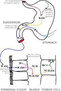
        • The vitB12 Journey.
          IF-Intrinsic Factor; Cbl-vitB12/ or Cobalamin; TC II-transcobalamin II)
        • Testing
          • Lack of gastric HCl is an indirect measure of lack of IF as it is due to atrophy of gastric cells
          • Look for IF Ab's (Blocking-prevents B12 binding to IF; Binding-prevents B12-IF complex binding to the ileum). 70% of PA sufferers have these Ab's
          • If negative (30%) do a Schilling Test.
            • Patient's vitB12 binding proteins are saturated by giving a large dose of unlabelled B12 intravenously
            • Absorption (via the ileum) of cobalamin is measured following ingestion of radioactively labelled B12.
            • In a normal system, more than 10% of the radioactive cobalamin will be excreted in the urine over 24hours.
            • If less than 10% is excreted, the patient is given cobalamin + IF. If the patient is suffering from PA, greater than 10% of the labelled cobalamin will appear in the urine within 24hours, otherwise the patient can be said to have an absorption problem.
            • Low results may occur in patient's with kidney problems
    • Terminal ileal disease
      • Ileal resections
      • Ulcerative colitis (may be autoimmune)
      • Tropical sprue (generalised malabsorption syndrome - seen in New Guinea)
    • Competitive parasites
      • Diphylobothrium latum
    • Pancreatic failure (enzymes from the pancreas are required to allow binding of IF to vitB12)
    • Drug induced
    • Bacterial overgrowth (competition for vitB12)
  • Increased Requirement for B12
    • Pregnancy
    • Infancy
    • Cancer
    • Haemolytic anaemia
    • Leukaemia
    • Exfoliative dermatitis
    • Multiple myeloma
  • Disorders of Transport and Storage
    Folic Acid Deficiency
  • Folate is obtained from leafy vegetables
  • Deficiency results in anaemia in approx. 20 weeks
  • Require 50-100ug/day
  • Serum folate assay can be variable and should be carried out with a RBC folate assay (which indicates folate levels at the time of production of the RBCs)
  • Polyglutamate folate is the most common form of folate in food. The broken down, monoglutamate is transported across the intestinal mucosa, mostly at the jejunum and is primarily stored in the liver (capacity of 5-10mg)
    Condition
    Serum B12 Assay
    RBC Folate Assay
    Pernicious Anaemia
    Low
    Decreased
    Alcoholism
    Normal
    Low
    Acute Folate Deficiency
    Normal
    Normal

  • Folate is taken into cells as N5-methyl-tetrahydrofolate(THF). This must be demethylated (which is mediated by cobalamin) before conjugation can occur. If N5-THF is not conjugated, it will leak out of the cell again.
  • If cobalamin is in short supply, demethylation is impaired
  • Normally, dUMP is entirely converted to dTMP. With decreased levels of folate, this conversion is limited and a build-up of dUMP occurs instead. As a consequence, dUTP concentration rises and begins to replace dTTP in DNA synthesis.
  • Error correction attempts to replace the U's but fails due to the lack of T's and DNA synthesis is interrupted
  • Note: Goat's milk is lower in folate than cow's milk

    Causes

  • Nutritional
    • An incomplete diet is the most common cause
    • Alcoholics
    • Elderly - overcooking destroys folate
  • Malabsorption
    • Tropical (general bowel disruption) and non-tropical (densitive to gluten - coeliac disease) sprue
    • Alcohol - interferes with absorption
    • Drug related - convulsive drugs, folate antagonists (eg. methotrexate-used for treatment of some neoplastic diseases), oral contraceptives
    • Anatomic and functional intestinal abnormalities
  • Increased Folate Requirement
    • Pregnancy
    • Infancy
    • Haemolytic anaemia
    • Acute leukaemia
    • Exfoliative dermatitis
    • Multiple myeloma
    • Metastatic cancer
    • Haemodialysis
  • Defective Utilization of Folate
  • Enzyme Deficiency

Sunday, 11 December 2011

Hematology Medicine mcqs

Hematology Medicine mcqs of previous year prepg exam

Download Hematology Medicine mcq

Breast Surgery theory & mcqs

very good notes of breast surgery and also contain good mcqs
Download Breast Surgery theory & mcqs

Guidelines for diagnosis and treatment of malaria in india

Poliomyelitis

it is very short ppt presentation of poliomyelitis
Download poliomyelitis ppt

MCQs in Clinical Pharmacy

A book in pdf formate contain many question of clinical pharmacy

Download MCQs in Clinical Pharmacy book (pdf)

Healthy Weight Loss Strategies

Sunday, 4 December 2011

Bronchial asthma


Bronchial asthma mcqs are in ppt formate
             these que. are asked in prepg exams

            https://rapidshare.com/files/3341845272/Asthma_mcqs.pptx

(for download right click on link and open in new tab then download